Why MRI findings, rotator cuff “damage,” and bursitis still don’t explain most shoulder pain
First published 2014 · Updated and re-released 2026
Shoulder pain remains one of the most over-imaged and over-operated musculoskeletal conditions in modern healthcare. Despite over a decade of accumulating evidence, many patients are still told that their pain is caused by “tears,” “bursitis,” or “impingement” seen on MRI, findings that, in most cases, are poorly correlated with symptoms.
This article revisits the evidence, much of which has only strengthened since it was first written, and outlines why exercise-based rehabilitation, not surgery, remains the treatment of choice for most shoulder pain.
MRI findings and shoulder pain: correlation is not causation
Rotator cuff tears:
The evidence is clear and consistent: rotator cuff tears are extremely common in people without shoulder pain.
Large imaging studies have repeatedly shown high rates of partial and full-thickness rotator cuff tears in asymptomatic individuals, including overhead athletes.
Many of these individuals retain full strength, function, and sporting participation for years without treatment.
Tear size does not reliably predict pain, disability, or prognosis.
Even in people who do have shoulder pain, symptoms frequently resolve with conservative care — despite the tear remaining present on imaging.
The unavoidable conclusion is that MRI cannot determine whether a rotator cuff tear is the cause of a patient’s pain. Using the presence of a tear as a primary justification for surgery is therefore scientifically indefensible in most cases.
This helps explain a persistent paradox:
There is no clear consensus on indications for rotator cuff surgery, yet tens of thousands of repairs are performed annually.
Bursitis: a misleading label
“Bursitis” is another common MRI finding used to explain shoulder pain, and another poor explanation.
Imaging studies demonstrate bursal changes in nearly all asymptomatic shoulders.
Bursitis-like MRI appearances are routinely seen after rotator cuff repair, even in patients with no pain or functional limitation.
Comparative studies show no meaningful association between bursal signal changes and symptoms.
In short:
Bursitis on MRI is usually an incidental finding, not a diagnosis.
The overlooked problem: how MRI results change pain
One of the most clinically important, and still under-appreciated issues is what happens after scan results are communicated.
Pain is not a direct read-out of tissue damage. It is shaped by expectation, threat perception, anxiety, and meaning.
When patients are told they have:
“tears”
“degeneration”
“damage”
“wear and tear”
they may understandably begin to see themselves as structurally broken, fragile, or in need of repair. This can:
Increase fear and movement avoidance
Reduce confidence and activity
Narrow behaviour in progressively smaller “safe” circles
Promote chronicity
Neuroscience research shows that expectations alone can amplify pain-related brain activity, particularly in prefrontal and limbic regions involved in threat prediction. Negative expectations can also trigger anxiety-driven biochemical processes that facilitate pain transmission.
For patients with pre-existing anxiety (common in persistent pain) this nocebo effect is particularly potent.
In practical terms, poorly framed MRI results can worsen pain, not explain it.
Surgery for “impingement”: still no advantage over exercise
High-quality trials and long-term follow-ups have consistently shown:
Arthroscopic decompression and acromioplasty provide no clinically meaningful benefit over structured exercise
Surgical treatment is not cost-effective
Long-term outcomes are equivalent, or no better, than conservative care
This mirrors findings from multiple sham-surgery trials across medicine (including specifically for the shoulder), where patients undergoing simulated surgery often improve just as much as those receiving the real procedure.
The key question, then, is not whether some patients feel better after surgery - but why.
The ritual, meaning, and expectation surrounding surgery are powerful. Without adequate controls for these effects, mechanical explanations for surgical benefit become increasingly implausible.
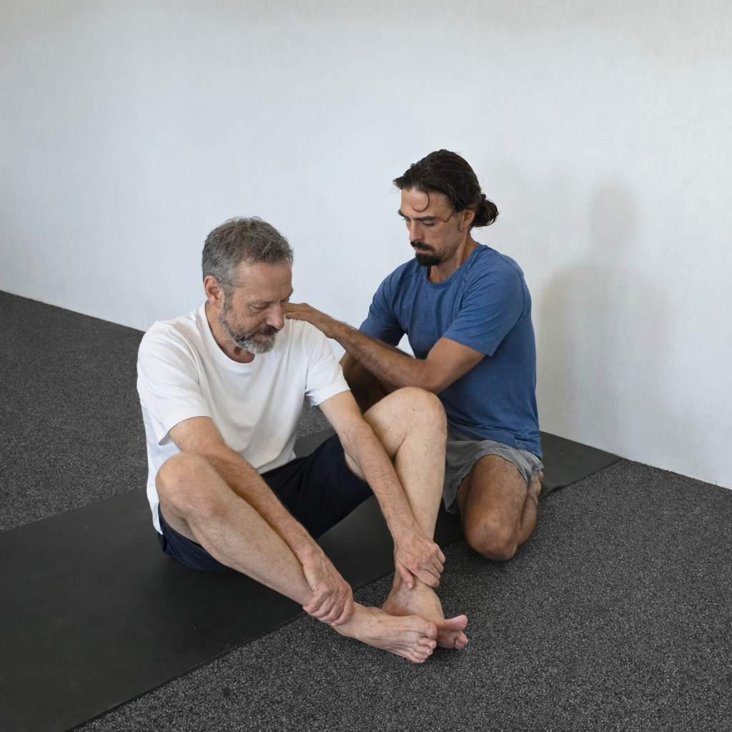
What actually works: exercise, education, and nervous system regulation
The most robust evidence supports individualised, supervised exercise rehabilitation as first-line care for shoulder pain, including in the presence of imaging “abnormalities.”
Effective programs typically include:
Progressive shoulder loading
Proprioceptive and neuromuscular training
Gradual exposure to feared or avoided movements
Crucially, outcomes improve further when rehabilitation also addresses:
Anxiety and threat sensitivity
Breathing patterns
Autonomic regulation
Cognitive and perceptual aspects of pain
This is where neurocognitive exercise approaches, including yoga-based therapy, are particularly valuable. Evidence suggests such approaches can reduce anxiety, improve motor control, and modulate central pain processing, with benefits maintained well beyond the treatment period.
Summary
Twelve years on, the core message remains unchanged, and better supported than ever:
MRI findings such as rotator cuff tears and bursitis rarely explain chronic shoulder pain
Surgery for non-traumatic shoulder pain offers no clear advantage over high-quality rehabilitation
Poorly communicated imaging results can worsen pain through nocebo mechanisms
Integrated, individualised exercise, addressing graduated loading and psychological variables, remains the most evidence-based approach
In this context, personalised yoga therapy is not an “alternative” treatment, but a clinically rational tool grounded in modern pain science, movement physiology, and neuroscience.
So what’s changed since 2014?
If anything, the last decade has strengthened, not weakened, the core arguments made in the original 2014 article.
1. “Impingement” has largely fallen out of favour
The idea that shoulder pain is caused by the rotator cuff being mechanically “pinched” under the acromion is now widely questioned. Contemporary models emphasise load tolerance, movement variability, and pain processing, rather than structural compression as a primary driver of symptoms.
2. MRI findings are now widely recognised as age-related and incidental
Large population studies and clinical guidelines increasingly acknowledge that findings such as:
rotator cuff tears
tendinopathy
bursal signal changes
are common, often asymptomatic, and poorly correlated with pain or disability — particularly in middle-aged and older adults. Imaging is now more often reserved for specific red flags, trauma, or surgical planning rather than routine diagnosis.
3. Sham-surgery evidence has become unavoidable
Since 2014, sham-controlled trials in orthopaedics (including meniscal and shoulder procedures) have further eroded the assumption that surgical “correction” of structural findings reliably improves pain. This has accelerated a broader reassessment of procedure-driven care for non-traumatic musculoskeletal pain.
4. Pain science has moved decisively toward a biopsychosocial model
Modern pain science now clearly frames pain as an output of the nervous system, influenced by context, expectation, threat perception, and prior experience — not a direct measure of tissue damage. The nocebo effects of alarming diagnostic language are now openly discussed in clinical education and guidelines.
5. Exercise remains first-line care — but the emphasis has evolved
While exercise was already supported in 2014, the emphasis has shifted from:
“correcting faulty structures”
to:
graded exposure, motor control, confidence, and nervous system regulation
Rehabilitation approaches that integrate movement, breathing, and cognitive safety, including psychology based and cognitive-functional exercise, now sit comfortably within mainstream evidence-informed care.
Bottom line:
The central claim of this article, that most shoulder pain is not a structural failure requiring repair, and that thoughtful, individualised rehabilitation outperforms surgery, has not been overturned. It has been validated.